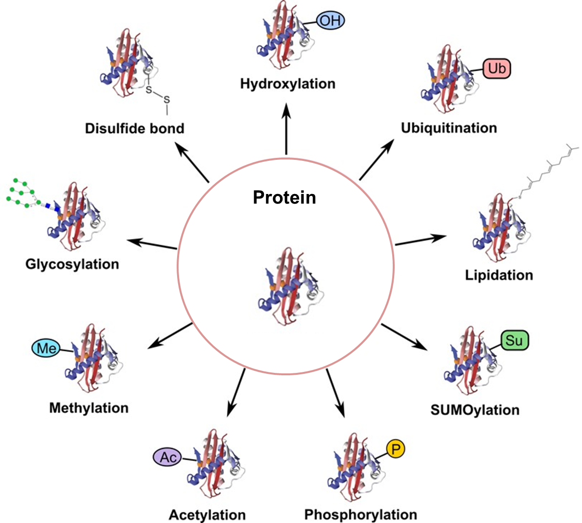
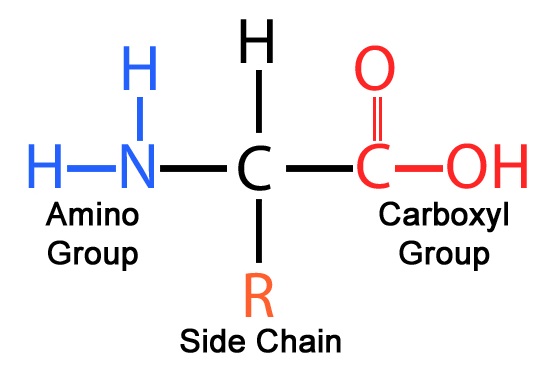
Molecular biology
  • Home
  • videos
    • Biology
    • Molecular Biology
    • Cell Biology
    • Biochemistry
    • Scientific Animation
    • Others
  • Books
    • Cell and Molecular Biology
    • Biochemistry and Biotechnology
    • Microbiology
    • Immunology
    • Biology
  • Notes
    • Molecular Biology
    • Biochemistry
    • Botechnology
  • Mock Test
Showing posts with label Molecular biology. Show all posts

Tuesday, August 6, 2024

DNA (Deoxyribonucleic Acid) Chapter-7

 August 06, 2024     Molecular biology     No comments   

 DNA Packaging: 

DNA packaging is a highly regulated process that enables the compaction of genetic material to fit within the confines of the cell nucleus while still allowing access for essential biological processes such as transcription, replication, and repair. This organization involves a hierarchical structure, from the basic nucleosome unit to higher-order chromatin structures.

1. Nucleosome Formation

Histones and DNA Interaction:

  • Histone Proteins: DNA is first wrapped around histone proteins to form the basic unit of chromatin, the nucleosome. Histones are small, highly basic proteins that neutralize the negative charge of DNA through electrostatic interactions.
  • Histone Octamer: A nucleosome core particle consists of an octamer of histone proteins (two each of H2A, H2B, H3, and H4). The DNA, approximately 146 base pairs in length, wraps around this octamer about 1.7 times, forming a histone core.
  • Histone H1: The linker histone H1 binds to the entry and exit sites of DNA on the nucleosome, stabilizing the nucleosome structure and promoting higher-order chromatin formation.
Reference: https://microbiologynotes.org/histones-types-and-its-functions/#google_vignette

Structural Characteristics:

  • Bead-on-a-String Model: The initial structure of chromatin is often described as a "bead-on-a-string" model, where nucleosomes (beads) are connected by linker DNA (string).

2. Formation of Higher-Order Chromatin Structures

30 nm Fiber:

  • Chromatin Fiber Formation: The "bead-on-a-string" nucleosomal array is further compacted into a 30 nm fiber. This fiber can be organized into two main models: the solenoid model (a helical structure) and the zigzag model (a less regular, more extended structure).
  • Compaction: The 30 nm fiber is believed to be stabilized by interactions between histone tails and linker DNA.

Loop Domains and Chromatin Organization:

  • Loop Domains: The 30 nm fiber is organized into looped domains anchored to a nuclear matrix or scaffold. This organization involves a network of structural maintenance of chromosomes (SMC) proteins, such as cohesin and condensin.
  • Chromosome Territories: In the nucleus, individual chromosomes occupy distinct territories. This organization influences gene expression and chromatin accessibility by reducing interactions between chromosomes.

3. Histone Modifications and Chromatin Remodeling

Post-Translational Modifications:

  • Acetylation: Histone acetylation, mediated by histone acetyltransferases (HATs), adds acetyl groups to lysine residues, reducing their positive charge and decreasing histone-DNA interactions. This modification is generally associated with gene activation.
  • Methylation: Histone methylation can either activate or repress gene expression depending on the context. Methylation of H3K4 is often associated with active transcription, while methylation of H3K9 and H3K27 is linked to gene repression.
Reference: https://www.researchgate.net

  • Phosphorylation: Phosphorylation of histone H3, particularly at serine 10, is associated with chromosome condensation during mitosis and is also involved in DNA damage response.

Chromatin Remodeling Complexes:

  • ATP-Dependent Remodeling: Chromatin remodeling complexes, such as SWI/SNF and ISWI, use ATP hydrolysis to reposition, eject, or restructure nucleosomes, thereby influencing chromatin accessibility and gene expression.

4. Chromatin Types: Euchromatin vs. Heterochromatin

Euchromatin:

  • Characteristics: Euchromatin is less densely packed and is associated with active gene transcription. It has a more open chromatin structure that allows access to the transcriptional machinery.
  • Role in Transcription: Euchromatin regions are generally rich in gene-dense areas and are frequently associated with active genes and regulatory elements.

Heterochromatin:

  • Characteristics: Heterochromatin is more compact and transcriptionally inactive. It can be classified into:
    • Constitutive Heterochromatin: Permanently compacted regions, such as centromeres and telomeres, involved in maintaining chromosome stability.
    • Facultative Heterochromatin: Can switch between an active and inactive state depending on cellular conditions. Examples include the X chromosome inactivation in females.
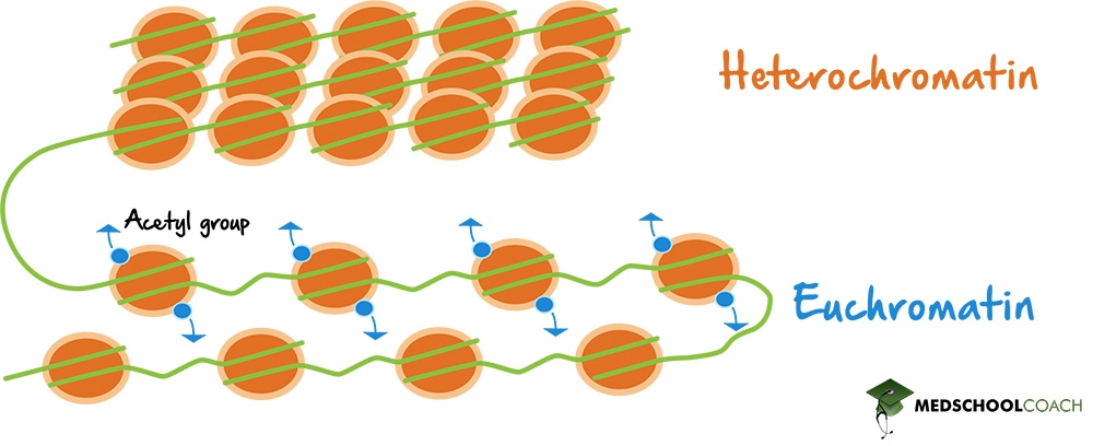
Reference: https://www.medschoolcoach.com/heterochromatin-vs-euchromatin-mcat-biology/

5. DNA Packaging in Prokaryotes

Nucleoid Region:

  • Organization: Prokaryotic DNA is organized in the nucleoid region, which lacks a defined membrane-bound nucleus. DNA is associated with various non-histone DNA-binding proteins.
  • Supercoiling: Prokaryotic DNA is typically negatively supercoiled, which introduces tension into the DNA, aiding in its compaction and playing a role in replication and transcription.

DNA Binding Proteins:

  • Structural Maintenance: Proteins such as HU (Histone like DNA binding proteins) and IHF (Integration host factors) help in organizing DNA into loops and domains within the nucleoid, affecting gene expression and DNA replication.


Reference: https://europepmc.org/article/med/32373086

6. Chromosome Condensation During Mitosis

Chromosome Structure:

  • Condensation: During cell division, chromatin undergoes further condensation to form distinct, visible chromosomes. This process is facilitated by condensin complexes, which help in the compaction of chromatin and ensure proper segregation of chromosomes.
  • Mitotic Chromosomes: The fully condensed mitotic chromosomes are highly compacted to ensure that DNA is accurately divided between daughter cells during mitosis.

Reference: https://f1000research.com/articles/5-1807

Functional Implications

Gene Regulation:

  • Transcriptional Control: DNA packaging affects the accessibility of genes for transcription. Modifications and higher-order structures can either promote or inhibit gene expression based on the cellular context.

Genomic Stability:

  • Chromosome Integrity: Proper DNA packaging is crucial for maintaining genomic stability. Errors in chromatin organization can lead to genomic instability and contribute to various diseases, including cancer.

Please, give your suggestions in comment section.

Follow our blogs for more updates and protocols: https://iammolecularbiologist.blogspot.com/ 

Follow us on WhatsApp: https://chat.whatsapp.com/I30OdffWlJhGABXsTmKo95

Read More
  • Share This:  
  •  WhatsApp
  •  Facebook
  •  Twitter
  •  Google+
  •  Stumble
  •  Digg

Thursday, August 1, 2024

Protein Chapter-4

 August 01, 2024     Molecular biology     No comments   

Protein Translocation

Overview of Protein Translocation

Protein translocation is a sophisticated and critical biological process involving the movement of proteins from their synthesis site to their specific destinations. It encompasses mechanisms like co-translational and post-translational translocation, and involves specialized molecular machines and signals.

2. Mechanisms of Protein Translocation

a. Co-translational Translocation

  1. Signal Sequence and SRP Recognition:

    • Signal Sequence: The signal sequence is an N-terminal peptide, often rich in hydrophobic residues, that directs the nascent polypeptide to the ER.
    • Signal Recognition Particle (SRP): SRP is a ribonucleoprotein complex consisting of an RNA component (SRP RNA) and six proteins (SRP54, SRP19, SRP68, SRP72, SRP9, SRP14). SRP54 binds to the signal sequence and the ribosome, pausing translation.
  2. SRP-Receptor Interaction:

    • Docking to ER Membrane: SRP binds to the SRP receptor (SR) on the ER membrane, which is composed of SRα and SRβ subunits. This interaction facilitates the transfer of the ribosome to the Sec61 translocon.
  3. Sec61 Translocon:

    • Structure: The Sec61 complex forms a protein-conducting channel in the ER membrane. It is composed of three main subunits (Sec61α, Sec61β, and Sec61γ) and associates with various auxiliary proteins such as TRAM (Translocon Associated Protein).
    • Function: The Sec61 channel opens to allow the polypeptide chain to enter the ER lumen. It also undergoes conformational changes to accommodate different stages of protein transport.
  4. Signal Peptide Cleavage and Protein Folding:

    • Signal Peptidase: The signal peptide is cleaved off by signal peptidase in the ER lumen. The mature protein then folds into its functional conformation with the aid of ER chaperones like BiP (Binding Immunoglobulin Protein), calnexin, and calreticulin.
  5. Quality Control:

    • ERAD (ER-Associated Degradation): Misfolded proteins are targeted for degradation by the ubiquitin-proteasome system. They are retro-translocated to the cytosol and tagged with ubiquitin for proteasomal degradation.
Reference: https://www.researchgate.net/figure/Schematic-representation-of-the-co-translational-translocation-of-a-polytopic-membrane_fig5_259675856

b. Post-Translational Translocation

  1. Mitochondrial Import:

    • Precursor Proteins: Mitochondrial proteins are synthesized with mitochondrial targeting sequences (MTS) that are recognized by the TOM (Translocase of the Outer Membrane) complex.
    • TOM Complex: TOM consists of multiple subunits, including TOM20 and TOM70 (receptors) and TOM40 (channel). It facilitates the initial import of precursor proteins into the intermembrane space.
    • TIM Complexes: TIM23 and TIM22 complexes mediate the translocation of proteins across the inner mitochondrial membrane. TIM23, in conjunction with the membrane potential, translocates proteins into the matrix, while TIM22 handles the insertion of proteins into the inner membrane.
Reference: https://www.researchgate.net/figure/Protein-translocation-a-Diagram-depicting-post-translational-translocation-into-the_fig2_343728797

  1. Nuclear Import:

    • Nuclear Localization Signal (NLS): Proteins destined for the nucleus contain NLS, which is recognized by importins. Importins are also known as karyopherins.
    • Nuclear Pore Complex (NPC): The NPC is a large structure composed of approximately 30 different nucleoporins (Nups). It forms a selective barrier that regulates the passage of proteins and RNAs between the nucleus and cytoplasm.
    • Importins and Ran Cycle: Importins bind to the NLS-containing cargo in the cytoplasm, transport it through the NPC, and release it in the nucleus. The GTPase Ran binds to importin in the nucleus, facilitating the release of the cargo and recycling of importins back to the cytoplasm.
  2. Chloroplast Import:

    • TOC Complex: The Translocon at the Outer Chloroplast membrane (TOC) complex consists of TOC75 (a channel-forming component) and TOC159/TOC33 (receptors).
    • TIC Complex: The Translocon at the Inner Chloroplast membrane (TIC) complex helps in the translocation of proteins across the inner membrane into the stroma. The TIC complex includes TIC110 and TIC40.


Reference: https://www.sciencedirect.com/science/article/pii/S0167488910000169

c. Vesicular Transport

  1. Vesicle Formation:

    • Coat Proteins: Vesicle formation involves coat proteins like clathrin (for transport between the Golgi and plasma membrane) and COPI/COPII (for ER-Golgi transport). Clathrin forms a triskelion shape and coats the vesicle, while COPI and COPII form coats on the cytoplasmic surface of the vesicle.
  2. Vesicle Transport:

    • Motor Proteins: Vesicles are transported along cytoskeletal tracks (microtubules) by motor proteins such as kinesins and dyneins. These motor proteins use ATP to move vesicles to their target membranes.
    • Anterograde transport refers to the movement of cargo from the endoplasmic reticulum (ER) towards the cell membrane or other destinations like the Golgi apparatus and lysosomes. This transport is essential for delivering newly synthesized proteins and lipids to their functional locations.
    • Retrograde transport involves the movement of cargo from the plasma membrane or Golgi back to the ER or other earlier compartments. This process is crucial for recycling components, retrieving misfolded proteins, and regulating cellular homeostasis.

Reference: https://www.researchgate.net/figure/Cytoplasmic-Dynein-and-Kinesin-Power-Axonal-Transport-Schematic-diagram-of-the_fig1_6784713

  1. Vesicle Fusion:

    • SNARE Proteins: The fusion of vesicles with target membranes is mediated by SNARE proteins. v-SNAREs (vesicle-SNAREs) on the vesicle and t-SNAREs (target-SNAREs) on the target membrane interact to facilitate membrane fusion and cargo release.
Reference: https://www.researchgate.net/figure/Basic-machinery-controlling-membrane-fusion-and-SNARE-recycling-SNAREs-and-tethers-are_fig3_51501699

3. Advanced Aspects of Protein Translocation

a. Signal Recognition and Integration:

  • Signal Sequences and Sorting Signals: Proteins contain various signal sequences and sorting signals that determine their final destination. The specificity of signal recognition by cellular machinery ensures correct protein localization.

b. Post-Translational Modifications:

  • Phosphorylation, Glycosylation, and Acetylation: These modifications can affect protein stability, localization, and function. For instance, phosphorylation of a nuclear localization signal can regulate nuclear import.

c. Diseases Related to Protein Translocation:

  • Alpha-1 Antitrypsin Deficiency: Caused by defective folding and translocation of the alpha-1 antitrypsin protein, leading to liver disease and lung damage.
  • Hereditary Spastic Paraplegia: Associated with mutations affecting protein import into mitochondria, resulting in neurodegenerative symptoms.

d. Experimental Techniques for Studying Protein Translocation:

  • Live-Cell Imaging: Techniques such as fluorescence recovery after photobleaching (FRAP) and fluorescence resonance energy transfer (FRET) help study dynamic aspects of protein localization and movement in real-time.
Reference: https://www.imperial.ac.uk/photonics/research/biophotonics/techniques/fluorescence-imaging-and-metrology/forster-resonant-energy-transfer-fret/

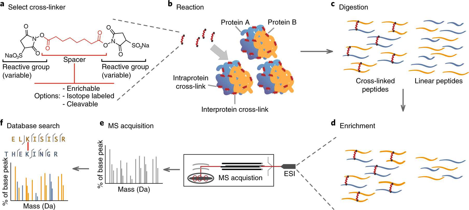
  • Cross-Linking and Mass Spectrometry: These methods are used to study protein-protein interactions and the identification of transient protein complexes involved in translocation.
Reference: https://www.nature.com/articles/s41594-018-0147-0


Please, give your suggetions in comment section.

Follow our blogs for more updates and protocols: https://iammolecularbiologist.blogspot.com/ 

Follow us on WhatsApp: https://chat.whatsapp.com/I30OdffWlJhGABXsTmKo95

Read More
  • Share This:  
  •  WhatsApp
  •  Facebook
  •  Twitter
  •  Google+
  •  Stumble
  •  Digg

Monday, July 29, 2024

RNA (Ribonucleic acid) Chapter-3

 July 29, 2024     Molecular biology     No comments   

RNA Splicing

1. Introduction to RNA Splicing

  • Definition: RNA splicing is a post-transcriptional modification process in eukaryotes where introns (non-coding regions) are removed from a pre-messenger RNA (pre-mRNA) transcript and exons (coding regions) are joined together to produce a mature mRNA molecule. This mature mRNA can then be translated into a protein.

  • RNA splicing is a fundamental cellular process in eukaryotes that transforms primary RNA transcripts into functional messenger RNAs (mRNAs). This process is essential for gene expression, allowing cells to produce a wide variety of proteins from a single gene through the selective removal of non-coding sequences and the joining of coding sequences.

  • Importance: Essential for the production of functional mRNA that can be translated into protein. It also allows for the generation of multiple protein isoforms from a single gene through alternative splicing.

2. Components Involved in Splicing

a. Pre-mRNA:

  • Introns: Intervening sequences that are transcribed but not translated. They often contain regulatory elements and can range in size from a few nucleotides to several kilobases.
  • Exons: Coding sequences that are retained in the mature mRNA. They are the segments that are expressed as proteins.

b. Splice Sites:

  • 5' Splice Site (Donor Site): Typically has a conserved GU sequence at the 5' end of the intron.
  • 3' Splice Site (Acceptor Site): Typically has a conserved AG sequence at the 3' end of the intron, often preceded by a pyrimidine-rich tract known as the polypyrimidine tract.
  • Branch Point: Located within the intron, usually 18-40 nucleotides upstream of the 3' splice site, and characterized by a conserved adenine residue that plays a crucial role in the splicing reaction.

c. Spliceosome:

  • Definition: A large, multi-component molecular machine composed of small nuclear ribonucleoproteins (snRNPs) and numerous associated proteins that carry out splicing.
  • Major snRNPs:
    • U1 snRNP: Recognizes and binds to the 5' splice site.
    • U2 snRNP: Binds to the branch point, creating a branch point complex.
    • U4/U6 snRNPs: Form a complex that stabilizes the spliceosome and assists in the catalytic steps.
    • U5 snRNP: Brings the 5' and 3' splice sites together and facilitates exon ligation.

3. Splicing Mechanism

a. Spliceosome Assembly:

  1. Initial Recognition:

    • U1 snRNP binds to the 5' splice site of the intron, establishing the early complex.
    • U2 snRNP binds to the branch point sequence, causing a conformational change that positions the branch point adenine for the first catalytic step.
  2. Complex Formation:

    • U4/U6 and U5 snRNPs join to form the mature spliceosome, which undergoes a series of conformational changes to become catalytically active.

b. Splicing Reaction:

  1. First Transesterification Reaction:
    • The 2' hydroxyl group of the branch point adenine attacks the 5' splice site, forming a lariat structure and producing a free 5' exon.
  2. Second Transesterification Reaction:
    • The 3' hydroxyl group of the 5' exon attacks the 3' splice site, leading to the ligation of the exons and release of the intron lariat.
Reference: https://www.researchgate.net 

c. Post-Splicing:

  • The lariat intron is debranched and degraded by cellular exonucleases and debranching enzymes.

4. Alternative Splicing

Definition: A process that allows a single gene to generate multiple mRNA isoforms by including or excluding specific exons or introns.

Types:

  • Exon Skipping: Exons are selectively excluded from the final mRNA, resulting in different protein variants.
  • Mutually Exclusive Exons: Only one of a pair or group of exons is included in the final mRNA, providing protein diversity.
  • Intron Retention: Retaining some introns in the mature mRNA can alter protein function or regulation.
  • Alternative 5' or 3' Splice Sites: Variation in splice site usage results in different mRNA isoforms.
Reference: https://en.wikipedia.org/wiki/Alternative_splicing

Significance: Increases proteomic diversity and allows cells to adapt to different developmental stages or environmental conditions.

5. Regulation of Splicing

a. Splicing Factors:

  • Splicing Activators:
    • SR Proteins (Serine/Arginine-rich proteins): Bind to exonic splicing enhancers (ESEs) and promote the inclusion of exons.
  • Splicing Repressors:
    • hnRNPs (Heterogeneous Nuclear Ribonucleoproteins): Bind to exonic splicing silencers (ESSs) and inhibit splicing.
Reference: https://www.mdpi.com/2073-4425/13/9/1659

b. Regulatory Sequences:

  • Exonic Splicing Enhancers (ESEs): Sequences within exons that enhance splicing by recruiting splicing activators.
  • Exonic Splicing Silencers (ESSs): Sequences within exons that inhibit splicing by recruiting splicing repressors.

c. Cellular Context:

  • Splicing is regulated by cell-type-specific splicing factors, cellular signals, and developmental cues. Splicing can be modulated in response to stress, signaling pathways, and developmental stages.

6. Clinical Implications

a. Splicing Mutations:

  • Cancer: Aberrant splicing can lead to the production of oncoproteins or loss of tumor suppressor proteins, contributing to cancer progression.
  • Genetic Disorders: Splicing mutations can cause diseases such as:
    • Spinal Muscular Atrophy (SMA): Caused by mutations affecting the SMN1 gene splicing.
    • Thalassemia: Caused by mutations that disrupt the splicing of β-globin mRNA.

b. Therapeutic Approaches:

  • Antisense Oligonucleotides (ASOs): Target specific splice sites to correct splicing defects in genetic disorders (e.g., nusinersen for SMA).
  • Small Molecules: Modulate splicing by interacting with splicing factors or the spliceosome (e.g., drugs targeting spliceosomal components in cancer).

7. Techniques and Tools

a. RNA Sequencing (RNA-seq):

  • Purpose: Provides a comprehensive view of transcriptome expression, including splicing patterns and alternative splicing events.
  • Applications: Used for gene expression analysis, discovery of novel splicing events, and understanding splicing regulation.

b. Splicing Reporter Assays:

  • Purpose: Assess the impact of specific splicing elements, mutations, or treatments on splicing efficiency and accuracy.

c. Computational Tools:

  • Splicing Prediction Algorithms: Tools like SpliceSiteFinder, MaxEntScan, and other bioinformatics software predict splice sites and potential alternative splicing events based on sequence data

Please, give your suggetions in comment section.

Follow our blogs for more updates and protocols: https://iammolecularbiologist.blogspot.com/ 

Follow us on WhatsApp: https://chat.whatsapp.com/I30OdffWlJhGABXsTmKo95
Read More
  • Share This:  
  •  WhatsApp
  •  Facebook
  •  Twitter
  •  Google+
  •  Stumble
  •  Digg

Protein Chapter-3

 July 29, 2024     Molecular biology     No comments   

Protein Function

Proteins are fundamental macromolecules composed of one or more long chains of amino acids. They are crucial for virtually all biological processes and serve a myriad of functions within cells and organisms. Each protein's function is determined by its unique sequence of amino acids and the specific three-dimensional structure it adopts. Here’s a breakdown of the major functional categories of proteins:

  1. Enzymatic Function: Proteins that act as enzymes facilitate biochemical reactions, increasing the rate at which reactions occur without being consumed in the process.

  2. Structural Support: Proteins provide physical support and shape to cells and tissues, contributing to the organization and integrity of biological structures.

  3. Transport: Proteins are involved in the movement of molecules across cell membranes and throughout the body, including the transport of gases, nutrients, and other essential substances.

  4. Signaling: Proteins play a key role in cellular communication, transmitting signals between cells and coordinating physiological processes through signaling pathways.

  5. Immune Response: Proteins are essential components of the immune system, defending the body against pathogens and foreign substances.

  6. Movement: Proteins are involved in cellular and organismal movement, including muscle contraction and intracellular transport.

  7. Storage: Proteins can store vital nutrients and energy sources for later use, playing a role in metabolism and homeostasis.

  8. Regulation: Proteins regulate various cellular processes, including gene expression and cell cycle progression, ensuring proper function and adaptation to environmental changes.

  9. Cell Recognition and Adhesion: Proteins mediate interactions between cells and between cells and their surroundings, facilitating tissue formation and communication.

Detailed Overview of Protein Functions

1. Enzymatic Function

Enzymes: Enzymes are a specific type of protein that acts as a biological catalyst. This means they speed up chemical reactions in the body without being consumed or permanently altered in the process. Enzymes are crucial for metabolic processes, such as digestion and cellular respiration.
  • Catalysts: Enzymes are specialized proteins that accelerate chemical reactions in the cell. They work by lowering the activation energy required for a reaction to proceed. The active site of an enzyme binds to the substrate, forming an enzyme-substrate complex. This interaction facilitates the conversion of substrates into products. For example, catalase breaks down hydrogen peroxide into water and oxygen, while lactase hydrolyzes lactose into glucose and galactose.

  • Regulation: Enzyme activity is finely tuned through mechanisms such as allosteric regulation, where molecules bind to sites other than the active site to modulate activity. Covalent modification, such as phosphorylation or acetylation, also affects enzyme function. Feedback inhibition occurs when the end product of a metabolic pathway inhibits an enzyme involved earlier in the pathway to prevent overproduction.


Reference:https://www.thoughtco.com/what-is-enzyme-structure-and-function-375555

2. Structural Support

  • Cytoskeleton: The cytoskeleton consists of protein filaments and tubules that provide structural support and shape to the cell. Key components include actin filaments, which form microfilaments that support cell shape and movement, and tubulin microtubules, which maintain cell shape, facilitate intracellular transport, and are involved in cell division.

  • Extracellular Matrix: The extracellular matrix (ECM) includes structural proteins like collagen and elastin. Collagen forms a fibrous network that provides tensile strength to tissues, while elastin imparts elasticity, allowing tissues to stretch and return to their original shape.

Reference: https://www.nature.com/scitable/topicpage/protein-function-14123348/

3. Transport
  • Molecular Transport: Proteins such as hemoglobin transport oxygen from the lungs to tissues and facilitate carbon dioxide transport from tissues back to the lungs. Hemoglobin’s quaternary structure (composed of four subunits) allows for cooperative binding of oxygen, increasing efficiency.

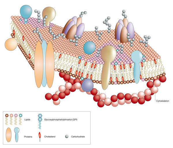
  • Cell Membrane Transport: Proteins in the cell membrane include channel proteins, which form pores allowing specific ions or molecules to pass through, and carrier proteins, which bind and transport substances across the membrane via conformational changes. For example, glucose transporters facilitate the uptake of glucose into cells.


Reference: https://www.researchgate.net

4. Signaling

  • Hormones: Protein hormones, such as insulin, regulate various physiological processes. Insulin binds to the insulin receptor, a receptor tyrosine kinase, initiating a signaling cascade that includes phosphoinositide 3-kinase (PI3K) and protein kinase B (AKT), leading to glucose uptake and metabolism.

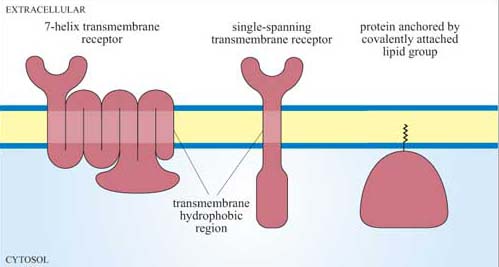
  • Receptors: Proteins like G-protein coupled receptors (GPCRs) are involved in signal transduction. Upon binding with a ligand, GPCRs activate G-proteins that modulate various intracellular pathways. For instance, beta-adrenergic receptors activate adenylyl cyclase, increasing cyclic AMP (cAMP) levels and activating protein kinase A (PKA).


Reference: https://www.open.edu/openlearn/science-maths-technology/cell-signalling/content-section-1.7


5. Immune Response
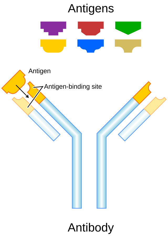
  • Antibodies: Immunoglobulins (Ig) are Y-shaped proteins with variable regions that specifically bind to antigens. They are crucial for identifying and neutralizing pathogens. The constant region of antibodies mediates immune responses such as complement activation and phagocytosis.

  • Complement Proteins: The complement system consists of plasma proteins that enhance the ability of antibodies and phagocytes to clear pathogens. Complement activation leads to the formation of the membrane attack complex (MAC), which forms pores in pathogen membranes and promotes their destruction.

Reference: https://en.wikipedia.org/wiki/Antibody

6. Movement
  • Muscle Contraction: Muscle contraction is driven by the interaction of actin and myosin filaments. The cross-bridge cycle involves myosin heads binding to actin, undergoing a conformational change (power stroke), and detaching after ATP hydrolysis, causing the filaments to slide past each other and shorten the muscle fiber.

  • Motility: Motor proteins such as kinesin and dynein transport cellular cargo along microtubules. Kinesin moves towards the plus end of microtubules (towards the cell periphery), while dynein moves towards the minus end (towards the cell center), using energy from ATP hydrolysis.

7. Storage

  • Nutrient Storage: Proteins like ferritin store iron in a non-toxic, bioavailable form. Ferritin’s protein shell encapsulates iron in a mineral core, which can be mobilized when needed, ensuring a supply of this essential nutrient.

  • Energy Storage: Proteins can be broken down into amino acids, which can be converted into glucose through gluconeogenesis or into ketone bodies through ketogenesis. This process is especially important during periods of fasting or prolonged exercise.

8. Regulation

  • Gene Expression: Transcription factors are proteins that regulate gene expression by binding to specific DNA sequences and modulating transcription. For instance, p53 is a transcription factor that activates genes involved in DNA repair and apoptosis in response to cellular damage.

  • Cell Cycle Control: Proteins like cyclins and cyclin-dependent kinases (CDKs) regulate the progression of the cell cycle. Cyclins bind to CDKs, forming complexes that phosphorylate target proteins, thereby driving the cell through various stages of the cell cycle.

Reference: https://pressbooks.bccampus.ca/humanbiology053/chapter/5-8-regulation-of-gene-expression/

9. Cell Recognition and Adhesion
  • Cell-Cell Recognition: Proteins such as selectins and integrins mediate cell-cell interactions and adhesion. Selectins bind to carbohydrate ligands on other cells, facilitating cell adhesion and migration, while integrins connect the cytoskeleton to the extracellular matrix, impacting cell movement and tissue formation.

  • Adhesion Molecules: Cadherins are involved in calcium-dependent cell-cell adhesion, forming structures like adherens junctions and desmosomes that maintain tissue integrity. Integrins facilitate cell-extracellular matrix interactions and play a role in cell signaling and tissue structure.


Please, give your suggetions in comment section.

Follow our blogs for more updates and protocols: https://iammolecularbiologist.blogspot.com/ 

Follow us on WhatsApp: https://chat.whatsapp.com/I30OdffWlJhGABXsTmKo95
Read More
  • Share This:  
  •  WhatsApp
  •  Facebook
  •  Twitter
  •  Google+
  •  Stumble
  •  Digg

Saturday, July 27, 2024

Protein Chapter-2

 July 27, 2024     Molecular biology     No comments   

Protein Translation

1. Introduction

Protein translation is a complex and highly regulated process by which cells convert the genetic code in mRNA into a specific polypeptide chain. This process involves the precise coordination of various molecular components and stages, ensuring that proteins are synthesized accurately and efficiently.

2. Components of Translation

  • mRNA (Messenger RNA):

    • Structure: Linear sequence with a 5' cap (7-methylguanylate cap) and a 3' poly(A) tail. The 5' cap protects the mRNA from degradation and aids in ribosome binding, while the poly(A) tail enhances stability and translation efficiency.
    • Coding Region: Contains coding sequences organized into codons, each specifying a single amino acid.

        Reference: https://www.geeksforgeeks.org/transfer-rna/
  • tRNA (Transfer RNA):

    • Structure: Cloverleaf secondary structure with an acceptor stem (where the amino acid attaches) and an anticodon loop (where the anticodon sequence binds to the mRNA codon).
    • Aminoacylation: tRNAs are charged with amino acids by aminoacyl-tRNA synthetases. This process involves the enzyme recognizing both the tRNA and the specific amino acid.
  • Ribosome:

    • Structure: Composed of two subunits. In prokaryotes, the small subunit is 30S and the large subunit is 50S, while in eukaryotes, the small subunit is 40S and the large subunit is 60S. The ribosome is a ribonucleoprotein complex that catalyzes peptide bond formation.
    • Functional Sites:
      • A (Aminoacyl) Site: Binds incoming aminoacyl-tRNA.
      • P (Peptidyl) Site: Holds the tRNA with the growing polypeptide chain.
      • E (Exit) Site: Where deacylated tRNA exits the ribosome.

       Reference:Bartleby.com
  • Aminoacyl-tRNA Synthetases:

    • Function: Catalyze the esterification of an amino acid to its corresponding tRNA. Each synthetase is specific to a single amino acid and its cognate tRNAs.
  • Translation Factors:

    • Initiation Factors (IFs in prokaryotes, eIFs in eukaryotes): Facilitate ribosome assembly and mRNA binding.
    • Elongation Factors (EFs in prokaryotes, eEFs in eukaryotes): Assist in the delivery of aminoacyl-tRNA to the ribosome and translocation of the ribosome along the mRNA.
    • Release Factors (RFs in prokaryotes, eRFs in eukaryotes): Recognize stop codons and facilitate the release of the newly synthesized polypeptide.
     

3. Stages of Translation

3.1. Initiation

  1. Formation of the Initiation Complex:

    • Prokaryotes:
      • The small ribosomal subunit (30S) binds to the mRNA at the Shine-Dalgarno sequence, which is complementary to a sequence in the 16S rRNA of the ribosome.
      • The initiator tRNA carrying formylmethionine (fMet) binds to the start codon (AUG) in the P site.
      • Initiation factors (IF1, IF2, and IF3) assist in this process.
    • Eukaryotes:
      • The small ribosomal subunit (40S) along with initiation factors (eIF1, eIF2, eIF3) binds to the 5' cap of the mRNA.
      • The pre-initiation complex scans the mRNA for the AUG start codon.
      • The initiator tRNA, charged with methionine, binds to the start codon, assisted by eIF5B.
  2. Assembly of the Ribosome:

    • The large ribosomal subunit (60S) joins the small subunit (40S), forming the functional 80S ribosome in eukaryotes, or the 70S ribosome in prokaryotes.
    • The initiator tRNA occupies the P site, while the A site is primed for the next aminoacyl-tRNA.
    
      Reference: https://www.sciencedirect.com/science/article/abs/pii/S0962892416300769

3.2. Elongation

  1. Codon Recognition:

    • Aminoacyl-tRNA enters the A site of the ribosome. The correct tRNA is selected based on the codon-anticodon pairing. This process is mediated by elongation factor EF-Tu (prokaryotes) or eEF1A (eukaryotes), which ensures accuracy by binding GTP.
  2. Peptide Bond Formation:

    • The ribosome’s peptidyl transferase center, a ribozyme activity of the 23S rRNA in prokaryotes or the 28S rRNA in eukaryotes, catalyzes the formation of a peptide bond between the amino group of the amino acid in the A site and the carboxyl group of the amino acid in the P site.
  3. Translocation:

    • The ribosome moves along the mRNA by one codon. This movement is facilitated by elongation factor EF-G (prokaryotes) or eEF2 (eukaryotes) and involves the hydrolysis of GTP.
    • The tRNA in the A site moves to the P site, and the tRNA in the P site moves to the E site before exiting the ribosome.

3.3. Termination

  1. Stop Codon Recognition:

    • When a stop codon (UAA, UAG, or UGA) reaches the A site, it is recognized by release factors (RF1 and RF2 in prokaryotes, eRF1 in eukaryotes).
    • Release factors bind to the stop codon and promote the hydrolysis of the ester bond between the polypeptide chain and the tRNA in the P site.
  2. Polypeptide Release:

    • The newly synthesized polypeptide is released into the cytoplasm or the endoplasmic reticulum, depending on the cell type and protein destination.
  3. Disassembly:

    • The ribosomal subunits dissociate from the mRNA, and the tRNA molecules are released.
    • The mRNA may be degraded or recycled for another round of translation.

  Reference:https://courses.lumenlearning.com/suny-mcc-microbiology/chapter/protein-synthesis-translation/

4. Post-Translational Modifications

  • Phosphorylation:

    • Enzymatic addition of phosphate groups to serine, threonine, or tyrosine residues by kinases. This modification can alter enzyme activity, protein-protein interactions, and cellular localization.
  • Glycosylation:

    • Addition of carbohydrate moieties to specific amino acid side chains, such as asparagine (N-linked glycosylation) or serine/threonine (O-linked glycosylation). Glycosylation affects protein folding, stability, and interactions.
  • Acetylation:

    • Addition of acetyl groups to lysine residues by acetyltransferases. Acetylation can influence gene expression, protein stability, and protein-protein interactions.
  • Cleavage:

    • Proteolytic cleavage by specific proteases can activate or inactivate proteins, remove signal sequences, or generate functional peptides from larger precursors.
   Reference: https://www.creative-proteomics.com/blog/index.php/strategies-for-post-translational-modifications-ptms/

5. Regulation of Translation

  • mRNA Stability:

    • Decay Mechanisms: mRNA stability is regulated by factors such as the presence of AU-rich elements (AREs) in the 3' UTR, which are bound by decay-promoting proteins.
    • Translational Repression: Binding of regulatory proteins or microRNAs (miRNAs) to the 3' UTR can inhibit translation or promote degradation.
  • Initiation Factors:

    • Phosphorylation: Phosphorylation of initiation factors (e.g., eIF2α) can inhibit translation initiation in response to stress or nutrient deprivation.
    • Regulatory Proteins: Specific proteins can modulate the activity of initiation factors and influence translation rates.
  • Global Regulation:

    • Stress Response: Cellular stress responses (e.g., heat shock, oxidative stress) can alter translation through pathways like the integrated stress response (ISR), which involves eIF2α phosphorylation and inhibition of global translation.
    • Nutrient Availability: Translation regulation in response to nutrient availability is mediated by signaling pathways such as the mTOR pathway, which affects the activity of translation initiation factors.

6. Translation in Prokaryotes vs. Eukaryotes

  • Prokaryotes:

    • Coupled Transcription and Translation: In prokaryotes, translation begins while the mRNA is still being synthesized (coupled transcription and translation).
    • Shine-Dalgarno Sequence: The small ribosomal subunit binds to the Shine-Dalgarno sequence in the mRNA, facilitating translation initiation.
  • Eukaryotes:

    • Spatial Separation: Transcription occurs in the nucleus, and translation occurs in the cytoplasm. mRNA undergoes processing (5' capping, splicing, and polyadenylation) before translation.
    • 5' Cap and Poly(A) Tail: The 5' cap and poly(A) tail enhance mRNA stability and translation efficiency. The cap-binding complex and poly(A)-binding proteins play key roles in initiation.

Please, give your suggetions in comment section.

Follow our blogs for more updates and protocols: https://iammolecularbiologist.blogspot.com/ 

Follow us on WhatsApp: https://chat.whatsapp.com/I30OdffWlJhGABXsTmKo95 
Read More
  • Share This:  
  •  WhatsApp
  •  Facebook
  •  Twitter
  •  Google+
  •  Stumble
  •  Digg

Thursday, July 25, 2024

Protein Chapter-1

 July 25, 2024     Molecular biology     No comments   

1. Introduction to Proteins

  • Definition: Proteins are macromolecules consisting of one or more long chains of amino acid residues.
  • Structure: Proteins are composed of carbon (C), hydrogen (H), oxygen (O), nitrogen (N), and occasionally sulfur (S) atoms. They are synthesized based on the genetic code.

2. Amino Acids

  • Basic Structure: Each amino acid contains:
    • A central alpha carbon (Cα)
    • An amino group (−NH2)
    • A carboxyl group (−COOH)
    • A hydrogen atom (H)
    • A distinctive side chain (R group), which determines the properties and functions of the amino acid.

         Reference: https://www.astrochem.org/sci/Amino_Acids.php
  • Classification: Amino acids are categorized based on the properties of their side chains:
    • Nonpolar, aliphatic: Glycine, alanine, valine, leucine, isoleucine, methionine.
    • Aromatic: Phenylalanine, tyrosine, tryptophan.
    • Polar, uncharged: Serine, threonine, cysteine, proline, asparagine, glutamine.
    • Positively charged (basic): Lysine, arginine, histidine.
    • Negatively charged (acidic): Aspartate, glutamate.

3. Protein Structure Levels

  • Primary Structure: The linear sequence of amino acids in a polypeptide chain, determined by mRNA.
  • Secondary Structure: Local folding patterns within the polypeptide stabilized by hydrogen bonds.
    • Alpha Helix: Right-handed coil stabilized by hydrogen bonds between the backbone carbonyl oxygen and amide hydrogen atoms four residues apart.
    • Beta Sheet: Strands connected laterally by at least two or three backbone hydrogen bonds, forming a sheet-like array.
  • Tertiary Structure: The overall three-dimensional shape of a polypeptide, stabilized by interactions between R groups, including hydrogen bonds, ionic bonds, hydrophobic interactions, and disulfide bridges (covalent bonds between cysteine residues).
  • Quaternary Structure: The structure formed by multiple polypeptide chains (subunits), which may be identical (homomers) or different (heteromers). Hemoglobin is an example of a protein with quaternary structure.

Reference:https://www.thoughtco.com/protein-structure-373563

4. Protein Functions

  • Enzymatic Activity: Catalysts that accelerate biochemical reactions by lowering activation energy. Examples include:
    • Hydrolases: Break bonds by hydrolysis (e.g., proteases).
    • Ligases: Join two molecules together (e.g., DNA ligase).
    • Oxidoreductases: Catalyze oxidation-reduction reactions (e.g., oxidases).
  • Structural Role: Provide cellular and tissue structure, as in:
    • Collagen: Found in connective tissues.
    • Keratin: Key component of hair, nails, and the epidermis.
  • Transport and Storage: Transport molecules or store ions and molecules.
    • Hemoglobin: Transports oxygen in the blood.
    • Ferritin: Stores iron in the liver.
  • Signal Transduction: Function as receptors and hormones in cellular communication.
    • Insulin: Regulates glucose metabolism.
    • G-protein-coupled receptors (GPCRs): Transmit signals across cell membranes.
  • Immune Response: Antibodies (immunoglobulins) identify and neutralize pathogens.

5. Protein Synthesis

  • Transcription: Synthesis of mRNA from a DNA template in the nucleus. Key steps include:
    • Initiation: RNA polymerase binds to the promoter region of the gene.
    • Elongation: RNA polymerase synthesizes mRNA by adding nucleotides complementary to the DNA template.
    • Termination: Transcription ends when RNA polymerase reaches a terminator sequence.
  • Translation: The mRNA is translated into a polypeptide chain at the ribosome.
    • Initiation: The ribosome assembles around the mRNA and the first tRNA.
    • Elongation: tRNAs bring amino acids to the ribosome, where they are added to the growing polypeptide chain.
    • Termination: The process ends when a stop codon is reached.

 Reference: https://www.geeksforgeeks.org/protein-synthesis-diagram/

6. Protein Digestion and Absorption

  • Digestion: Begins in the stomach with pepsin and continues in the small intestine with proteases (e.g., trypsin, chymotrypsin, carboxypeptidase).
  • Absorption: Amino acids and small peptides are absorbed by enterocytes in the small intestine via active transport and facilitated diffusion.

7. Protein Metabolism

  • Anabolism: Biosynthesis of proteins from amino acids.
    • Transamination: Transfer of an amino group from one amino acid to a keto acid, forming a new amino acid.
    • Aminoacyl-tRNA Synthetase: Enzyme that attaches amino acids to their corresponding tRNA.
  • Catabolism: Breakdown of proteins into amino acids and further into ammonia, urea, and keto acids.
    • Deamination: Removal of an amino group from an amino acid.
    • Urea Cycle: Converts toxic ammonia to urea for excretion.


  Reference:https://www.osmosis.org/notes/Protein_Metabolism

8. Protein Requirements and Sources

  • Daily Requirements: Varies by individual factors. The RDA is approximately 0.8 grams per kilogram of body weight for adults.
  • Sources:
    • High-Quality Proteins (Complete Proteins): Provide all essential amino acids in sufficient quantities (e.g., meat, eggs, dairy).
    • Lower-Quality Proteins (Incomplete Proteins): Lack one or more essential amino acids (e.g., most plant-based sources like grains, nuts, and legumes).

9. Protein Deficiency

  • Kwashiorkor: Severe protein deficiency characterized by edema, an enlarged liver, and dermatosis.
  • Marasmus: Severe calorie and protein deficiency, leading to significant wasting and stunted growth.

10. Protein Quality

  • Biological Value (BV): Measures the proportion of absorbed protein incorporated into body proteins. High BV proteins provide a greater proportion of utilized amino acids.
  • Protein Digestibility Corrected Amino Acid Score (PDCAAS): Evaluates protein quality based on amino acid requirements and digestibility.
  • Net Protein Utilization (NPU): Ratio of amino acids converted to proteins to the amino acids supplied.

11. Protein and Health

  • Muscle Maintenance and Growth: Essential for muscle protein synthesis, repair, and maintenance. Influenced by factors like exercise and protein intake.
  • Immune Function: Essential for the synthesis of immunoglobulins and other immune system components.
  • Hormone Production: Many hormones, such as insulin and growth hormone, are protein-based.

12. Protein Supplements

  • Types:
    • Whey Protein: Fast-digesting, high in essential amino acids, especially leucine.
    • Casein Protein: Slow-digesting, provides a steady release of amino acids.
    • Plant-Based Proteins: Derived from soy, pea, rice, etc., suitable for vegetarians and vegans.
  • Uses: Often used to supplement dietary protein intake for muscle building, recovery, and other health benefits.
Read More
  • Share This:  
  •  WhatsApp
  •  Facebook
  •  Twitter
  •  Google+
  •  Stumble
  •  Digg

Tuesday, July 23, 2024

RNA (Ribonucleic acid) Chapter-2

 July 23, 2024     Molecular biology     No comments   


RNA Transcription

1. Overview of RNA Transcription

RNA transcription is a highly regulated process that converts genetic information stored in DNA into RNA. This process is central to cellular functions, enabling the synthesis of proteins and other RNA molecules.

2. Key Components

  • DNA Template: The DNA template strand used for transcription is read in the 3' to 5' direction. The RNA polymerase synthesizes RNA in the 5' to 3' direction, meaning that RNA is complementary to the DNA template strand.

  • RNA Polymerase: This enzyme has multiple subunits and varies between prokaryotes and eukaryotes:

    • Prokaryotes: RNA polymerase is a multi-subunit complex with a core enzyme made up of two alpha subunits, one beta subunit, one beta-prime subunit, and a sigma factor that binds to the promoter.
    • Eukaryotes: RNA polymerase II, the enzyme responsible for mRNA synthesis, has 12 subunits and requires additional general transcription factors (GTFs) for promoter recognition and initiation.
  • Promoter: In prokaryotes, promoters typically have conserved sequences known as the -10 (Pribnow box) and -35 regions. In eukaryotes, the core promoter often includes a TATA box, located approximately 25-30 base pairs upstream of the transcription start site (TSS).

  • Terminators:

    • Prokaryotes: Two main types: rho-independent (intrinsic) and rho-dependent. Rho-independent terminators form a hairpin loop followed by a poly-U tract in the RNA that destabilizes the RNA-DNA hybrid.
    • Eukaryotes: Termination often involves cleavage and polyadenylation, where specific sequences (such as the poly-A signal) and associated factors lead to the cleavage of the pre-mRNA and the addition of the poly-A tail.

3. Transcription Process

a. Initiation

  1. Promoter Binding:

    • Prokaryotes: Sigma factors recognize specific promoter sequences and facilitate the binding of RNA polymerase.
    • Eukaryotes: General transcription factors (TFIID, TFIIA, TFIIB, etc.) bind to the promoter region and recruit RNA polymerase II.
  2. Formation of the Transcription Initiation Complex:

    • Prokaryotes: The RNA polymerase-sigma complex forms a closed complex with DNA, then transitions to an open complex where the DNA is unwound.
    • Eukaryotes: The pre-initiation complex (PIC) forms, including TFIID (which binds the TATA box), and RNA polymerase II is recruited. This process involves the formation of the transcription bubble.
  3. DNA Unwinding: The DNA double helix is locally unwound by RNA polymerase and helicase activity, creating a transcription bubble where RNA synthesis occurs.

b. Elongation

  1. RNA Synthesis: RNA polymerase synthesizes RNA by adding ribonucleotides complementary to the DNA template strand. The enzyme maintains a transcription bubble, where the RNA-DNA hybrid is about 8-9 base pairs long.

  2. Proofreading: RNA polymerase has a rudimentary proofreading mechanism known as pyrophosphorolysis, where misincorporated nucleotides are removed by backtracking and cleaving the RNA.


Reference: https://www.researchgate.net/figure/Transcription-is-a-highly-complex-process-subject-to-numerous-modes-of-regulation-A-A_fig1_315958971

c. Termination

  1. Termination Signal:

    • Prokaryotes: Rho-dependent termination involves the rho factor protein, which binds to the RNA and moves towards the RNA polymerase, facilitating its release.
    • Eukaryotes: The termination process involves cleavage of the pre-mRNA at a specific site and subsequent addition of the poly-A tail. The cleavage is mediated by endonucleases, and the poly-A tail is added by poly-A polymerase.

Reference:https://www.pinterest.com/pin/531002612290782860/
  1. RNA Release: After termination, RNA polymerase releases the RNA transcript and dissociates from the DNA.

  2. DNA Reformation: DNA re-anneals to restore the double helix structure, and the transcription bubble resolves.

4. Post-Transcriptional Modifications (in Eukaryotes)

  1. Capping:

    • 7-Methylguanylate (7mG) Cap: Added to the 5' end of the RNA molecule. The cap is essential for RNA stability, splicing, and translation. The addition involves a 5'-5' triphosphate bridge.
  2. Polyadenylation:

    • Poly-A Tail: Added to the 3' end of the RNA after cleavage. This modification increases mRNA stability and facilitates export from the nucleus. The tail is added by poly-A polymerase.
  3. Splicing:

    • Spliceosome: A complex of small nuclear RNAs (snRNAs) and proteins that carry out splicing. Splicing involves the removal of introns and the joining of exons through a two-step transesterification reaction.
    • Alternative Splicing: Allows for the production of multiple protein isoforms from a single gene by differentially including or excluding exons.

Reference: https://www.toppr.com/ask/question/describe-post-transcriptional-processing-of-rna-in-eukaryotes/

5. Types of RNA Produced

  • mRNA (Messenger RNA): Encodes the instructions for protein synthesis. Eukaryotic mRNAs have a 5' cap, coding sequence, and a poly-A tail. Prokaryotic mRNAs often lack a poly-A tail.

  • rRNA (Ribosomal RNA): Synthesized in the nucleolus and incorporated into ribosomes. rRNA is critical for ribosome structure and function. In eukaryotes, it includes 18S, 5.8S, 28S rRNAs, while prokaryotes have 16S and 23S rRNAs.

  • tRNA (Transfer RNA): Adaptor molecules that bring amino acids to the ribosome. Each tRNA has an anticodon that pairs with an mRNA codon and a corresponding amino acid attachment site.

  • Other Non-Coding RNAs: Includes microRNAs (miRNAs) and long non-coding RNAs (lncRNAs) involved in gene regulation, chromatin modification, and other cellular processes. miRNAs regulate gene expression by binding to complementary mRNA sequences, while lncRNAs can modulate gene expression through various mechanisms, including chromatin remodeling.

6. Regulation of Transcription

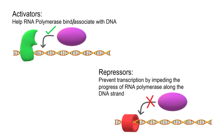
  • Transcription Factors: Proteins that regulate transcription by binding to specific DNA sequences. They can be activators (e.g., CREB, which binds to cAMP response elements) or repressors (e.g., REST, which represses neuronal gene expression in non-neuronal tissues).

  • Epigenetic Modifications:

    • DNA Methylation: Addition of methyl groups to cytosine residues in DNA, typically leading to gene silencing.
    • Histone Modifications: Post-translational modifications to histone proteins, including acetylation, methylation, and phosphorylation, that affect chromatin structure and gene accessibility.
  • Enhancers and Silencers:

    • Enhancers: DNA elements that can be located far from the gene they regulate. They increase transcriptional activity by interacting with the promoter through DNA looping.
    • Silencers: DNA elements that decrease transcriptional activity by binding repressor proteins and inhibiting transcription.

7. Significance

RNA transcription is essential for cellular function and organismal development. It allows for the dynamic regulation of gene expression in response to environmental cues and internal signals. Dysregulation of transcription can lead to diseases such as cancer, genetic disorders, and developmental abnormalities. Understanding the intricacies of transcription helps in the development of therapeutic strategies and provides insights into fundamental biological processes


Please, write your suggetions in comment section.

Follow our blogs for more updates and protocols: https://iammolecularbiologist.blogspot.com/  
Follow us on WhatsApp: https://chat.whatsapp.com/I30OdffWlJhGABXsTmKo95

Read More
  • Share This:  
  •  WhatsApp
  •  Facebook
  •  Twitter
  •  Google+
  •  Stumble
  •  Digg
Older Posts Home

Followers

Popular Posts

  • DNA (Deoxyribonucleic Acid) Chapter-7
      DNA Packaging:  DNA packaging is a highly regulated process that enables the compaction of genetic material to fit within the confines of ...
  • Basic Molecular Biology chapter -3
    Principles of gene regulation: Introduction to Gene Regulation Gene regulation is a critical process by which cells control the expression o...
  • RNA (Ribonucleic acid) Chapter-3
    RNA Splicing 1. Introduction to RNA Splicing Definition: RNA splicing is a post-transcriptional modification process in eukaryotes where in...
  • Mock Test on DNA Repair
    Start Quiz Submit Quiz Over! Repeat...

Categories

  • Biochemistry
  • MCQ
  • Mock test
  • Molecular biology
  • Molecular biology technology
  • Science

Blog Archive

  • ▼  2024 (30)
    • ▼  December (1)
      • Basic Molecular Biology chapter -3
    • ►  August (3)
    • ►  July (26)
  • ►  2022 (3)
    • ►  January (3)

Tags

Biochemistry MCQ Mock test Molecular biology Molecular biology technology Science

Featured post

Basic Molecular Biology chapter -3

Principles of gene regulation: Introduction to Gene Regulation Gene regulation is a critical process by which cells control the expression o...

Popular Posts

  • DNA (Deoxyribonucleic Acid) Chapter-7
      DNA Packaging:  DNA packaging is a highly regulated process that enables the compaction of genetic material to fit within the confines of ...
  • Basic Molecular Biology chapter -3
    Principles of gene regulation: Introduction to Gene Regulation Gene regulation is a critical process by which cells control the expression o...
  • Home

Copyright © Molecular biology | Powered by Blogger
Design by Hardeep Asrani | Blogger Theme by NewBloggerThemes.com | Distributed By Gooyaabi Templates